I just did a recent article for Midweek about pink eye.  Feel free to check it out – I just talk about prevention and treatment of conjunctivitis.  But, that’s not the topic of this post.  The Midweek writer’s first question to me was “What got you interested in pediatric ophthalmology?”.   I’m lucky enough that this question is such a non-brainer.  Is there anything better than restoring sight to a child? Complex surgery or a simple pair of glasses – both are so important in the developing vision of children.  That question made me think about one of our my first patients here in Hawaii.  I worked for a year after my fellowship as an attending at Boston Children’s Hospital. but there I was surrounded by senior physicians.  If I needed help on a case, or advice, they were there to assist.  When I moved out here with my husband 6 years ago, it was, honestly a bit terrifying.  I had lost my safety net and I had to build a practice from the ground up, somehow convincing parents that despite looking young and inexperienced, that I actually was a capable surgeon.

Niko was just a 6 year old boy when he came into see me my first month practicing at Honolulu Eye Clinic.

He had previously been living in California and had recently moved to Hawaii.  His mom had noted that he squinted a lot.   Niko was a smart little boy, but he had real trouble with the eye chart.  When I dilated his eyes, I discovered cataractsin both eyes.  A lot of people are often surprised that children and even babies can get cataracts.  It’s obviously much more rare than in adults, but it does occur.   I met with Niko and his sweet mom, Louise and told them that we should schedule surgery for Niko.

Marking Niko's eye for surgery in the pre-op area

This is what Niko’s mom wrote about her experience:

We first found out about a vision problem when Nikolas and I were reunited in Hawaii after a year of separation due to an illness of mine. He spent kindergarten year in San Jose California with my parents and siblings while I received treatment.While in San Jose he complained about not being able to see the board at school towards the end of the school year. When I picked him up and brought him back to Hawaii I decided to take him for a complete physical and suggested to the clinic physician for an eye exam as well.  They tested his eyes and we discovered that he couldn’t read most of the letters. The optometrist doing the exam referred us to Dr. Wong. He said that Niko may have cataracts. I was shocked and worried because he had just turned 6 yrs old.
 
Dr. Wong and her staff were so warm and welcoming. Even before we stepped into the clinic, I had already spoken to her on the phone several times. She told me about herself and her experience that gave me such relief to have found someone that can help my sons condition.  She performed surgery first on the right eye about 4 wks later and the left eye another 4 wks after that. The surgeries went well and Niko mentioned that everything so much brighter than before on the way to school a few days after. I was moved to tears of joy.
He now wears bifocal glasses that he loves and thrives in school. He is always excited to see Dr. Wong and her loving staff for regular check ups. He even mentioned to me several times that when he grows up he wants to become an eye doctor just like Dr. Wong so he can also help others see better.
 

Niko gave me this card.  Six years later, I still keep this card at my house.

After surgery, Niko’s first words to me were “I  can finally see the clock in school!”  His vision improved to 20/20 in each eye.

Each time he came for his post-operative visit, Niko would show off his latest dance moves for my staff.  I mean, seriously, is there anything better than getting that letter from a Mom or this card from a six year old?  He got cute bifocal glasses which he proudly wore all the time.

And, now 6 years later, Niko is an aspiring actor and model!


I know pediatric ophthalmology is not one of the “sexy” subspecialties of ophthalmology.  Everyone wants to do Cornea or Cataract with all the fun laser cataract machines or Retina with the cool new macular degeneration drugs that are coming out.  But, peds/strabismus has its HUGE rewards and I for one, cannot think of anything else I’d rather do.



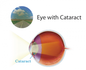
I’ll admit it – I eat gummy vitamins.  It’s the only way I can get myself to remember to take my prenatal vitamins and since I am nursing, it is extremely important for the nutrition of my child.  The beneficial effects of multi-vitamins are numerous – calcium for your bones, omega 3 fatty acids for neurologic development.   Patients routinely ask me if a multivitamin will help prevent the onset of cataracts.  Previous studies which have shown that vitamins can help prevent the progression of macular degeneration, never actually demonstrated that vitamins decrease the incidence of cataracts.  The Age Related Eye Disease Study (AREDS) showed that taking daily high doses of vitamins C and E, beta-carotene, zinc and copper can slow down the progress of age-related macular degeneration (AMD).   These are the vitamins that say “Eye Health” at Costco. One of my previous posts was about this protective effect of anti-oxidants and macular degneneration.  But, what about cataracts?  Well, first – what is a cataract?

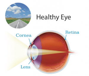
A cataract is a clouding of the normally clear lens within your eye, which results in blurred or distorted vision. The lens helps to focus light onto the retina, the part of the eye similar to the film in a camera.  Light enters the eye through the cornea, passes through the natural crystalline lens and is accurately focused onto the retina, providing a crisp, clear image.


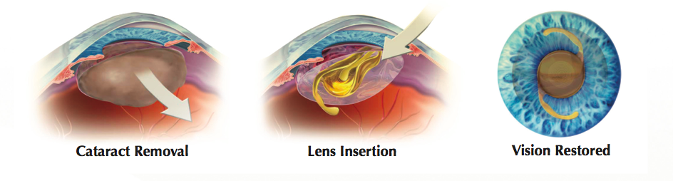
Cataracts are most often found in older patients but they can occur in people of any age. They can progress until eventually there is significant loss of vision, and neither diet, nor medications, will make the cataract go away. Surgery is required to improve vision.  Some people mistakenly think that a cataract is a growth in the eye and that surgery entails “cleaning the cataract out.”.  That’s not quite the case.  The cataract is removed with laser or ultrasound power and replaced with an artificial lens.

However, a new study examined a group of almost 15,000 male physicians , over the age of 50 and followed them over an average of 11 years.  The study was well designed, it was a prospective, double blind, randomized, placebo controlled trial, done at Brigham Women’s Hospital and Harvard Medical School. Half took a common daily multivitamin, as well as vitamin C, vitamin E and beta carotene supplements. The other half took a placebo.  Researches then followed the patients to assess if they developed cataracts or macular degeneration.  They found a roughly 9% decrease in incidence in the development of cataracts in the group who took the vitamins compared to placebo.  Nine percent may not seem like a lot, but when you think about the fact that over 22 million people in the United States alone have cataracts, that 9% translates to roughly 2 million people who may not get cataracts if taking these multivitamins.  On a world wide scale, it is estimated that cataracts cause 51% of blindness, affecting about 20 million people.  And, though cataracts are treatable, many people in developing countries do not have access to surgical care.  A simple multivitamin could make a huge difference in these lives, either delaying the onset or progression of cataracts


Many of you remember when I wanted to take “The Latisse Challenge” last year.  I was planning on using Latisse and taking weekly photographs of my lash growth.  But, since I was breast feeding my second son, I decided against doing it (Latisse is not FDA approved in pregnant or nursing moms).  And, since I got very quickly pregnant with Arya after ceasing nursing Taj, I really have never had the chance to try Latisse.  I have been jealously checking out  all of the people around me with their gorgeous eyelashes.  Even both my boys have lashes that Kim Kardashian would covet.


My Boys

To top it off, I have suffered from madarosis with pregnancy.  Madarosis is the medical term for loss of eyelashes and eyebrows.

There can be many different reasons for it – inflammation such as caused by infections, blepharitis, or even allergy.  Or it can be a sign of a systemic disease or condition, toxicity from medications, nutritional disorders, autoimmune disorder (lupus), tumors, hyperthyroidism or hypothyroidism, or traction (did you know that a lash curler can cause lash loss??).  For me, I think it’s been a combination of hormones and my lash curler.  The skimpier my lashes got, the more I tried to make every lash count – so I started curling them which really made things worse.  Oh, and I’m a notorious eye rubber.  I get bad allergies and I never remember to take my Pataday drops and just end up pulling and stretching my eyelid skin, which is never a good thing.

All of this lead me to try lash extensions.  Several of my friends have them and they are super popular here in Hawaii.  Many of my patients ask me about them and I wanted to try it out.  The licensing for lash extensions varies state to state.  In Hawaii, an aesthetician or cosmetologist can be licensed to apply lash extensions, even currently no classes or lectures are taught about lashes in these two fields.  Therefore, anyone can do lashes and you should make sure that the person you choose for your lash extensions does not skimp on the type glue, types of lashes and is diligent with their application.  After all, this is a non-surgeon using crazy glue and sharp instruments approximately 1 mm from your eye!  The American Academy of Ophthalmology cautions about the dangers of lash extensions.  They warn about:

  • Infection of the cornea
  • Infection or swelling of the eyelid
  • Permanent or temporary loss of lashes

I went to a lady who was highly recommended.  The process of lash extensions involves gluing, with cyanoacrylate glue (Dermabond – the same type of skin glue doctors sometimes use instead of stitches), lashes on to the base of your natural lashes.  I have since learned that there are a few different types of lashes – synthetic, silk and mink.  For mink lashes, think the Kardashians or Beyonce.

The adhesive should not be adherent to your eyelid skin.  The first time I had the lashes done, I liked their look and did not have difficulty with them.

The typical lash cycle consists of 4 different phases, and most women have between 100-200 upper lashes on each eye.


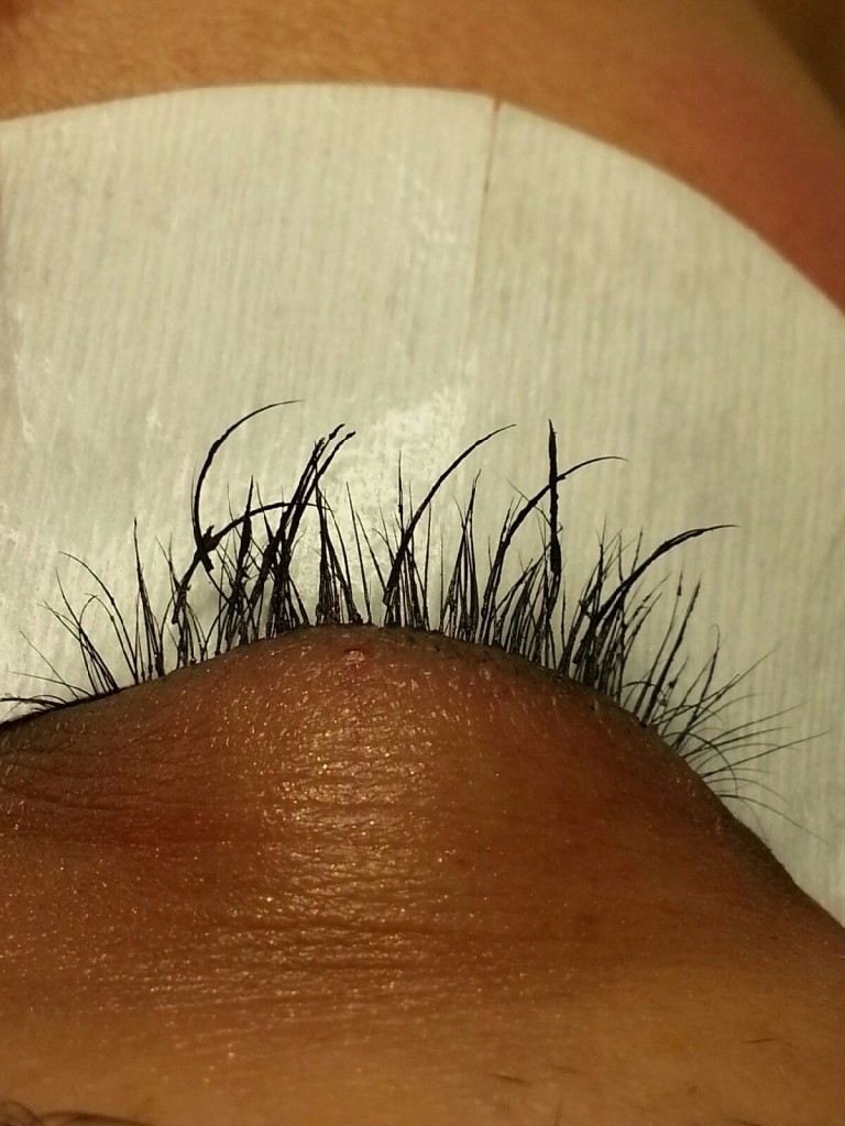
Therefore, even though it takes a full 3 months to cycle through your lashes, most women will want to get their lash extensions filled in every 3-4 weeks.  I went back for my fill=in and asked for more noticeable lashes.  One thing to note is that I had my baby 3 months ago.  This is important because, as many of you mamas out there know, like clockwork, right around the 3-4 month mark, all that luscious hair on your head that the baby hormones were promoting starts to fall out.  Same is true for your lashes.  Unfortunately, the lash lady saw that I had baby fine lashes, since I was shedding a lot of lashes due to the hormones, and applied extra glue to make the lashes stick better.  The fumes from the glue caused my eyes to tear throughout the entire process. The end result was that the lashes were too heavy and caused most of my real lashes to fall out.  Even though I’m an ophthalmologist, it didn’t occur to me to check the ingredients of the glue used.


Lash extensions glued to multiple of my lashes with excess glue at base

I looked online for studies regarding lash growth and lash extensions.  You might be scared yourself to try lash extensions because you’ve heard “it causes your lashes to fall out”.  In reality, no study has actually been performed to support or deny this assertion.  But, there are risks associated with lash extensions – irritation, inflammation, infection, allergic reaction and even madarosis.  Some of you may remember Kristin Chenowith showing up on David Leterman wearing huge sunglasses because of an allergic reaction from her lash extensions.


Long and short of it, I started developing irritation and redness of my upper eyelid skin from the lash extensions – I wanted them off.  I almost attempted to pull them off myself, but I knew that would result in more missing lashes.

So, I tried another lash stylist (is that what they are called?) who had been highly recommended by another friend.  Kristin Wood of the Kristin Wood salon.  She spent an hour and a half carefully removing all of the excess glue at the base of my lashes and the few errant lashes remaining.  At the conclusion, my upper lids were swollen and red and she advised me to wait  to get new lash extensions until my eyelids had healed.

Redness and swelling at base of lashes after lash removal

I knew that was the right call, so I came back in two weeks and she worked her magic.

New lashes!


One week later, only two of my lashes have fallen out.  The lashes are longer and feel much lighter than my previous ones.  When I touch them, they don’t feel stiff, but soft like my own lashes.  However, most people in the lash industry will admit that lash extensions do cause your natural lashes to fall out more quickly.  So, what should you do to avoid complications from lash extensions?  The FDA and American Academy of Ophthalmology offers the following advice:

  • Check the ingredients of the glue to make sure you are not allergic to it
  • If you have an eye infection or the skin around the eyes is inflamed, avoid lash extensions (as I did initially)
  • Make sure that the technician applying your lashes wears gloves and practices proper hygiene
  • Ensure that the aesthetician is properly certified and working at a reputable place.

If you develop an infection from the lash extensions, resist the urge to pull them out yourself.  Go to an ophthalmologist for treatment. An ophthalmologist can prescribe an antibiotic or antiobiotic/steroid ointment.  The lashes will fall out over the period of about six weeks and with it, the glue should also fall out in that time.

Also, beware of a lash stylist who tells you not to get your lashes wet at all.  Usually, you shouldn’t get them wet the first 24 hours, but after this, you should clean your lashes and remove make-up with an oil-free make-up remover.  Kristin advised to perform the same baby shampoo lid scrubs that I recommend to my patients with blepharitis to prevent build-up of protein and oil (and this was before she knew I was an ophthalmologist!)

So, what do I think of lash extensions?  I had one experience which seems to echo everything that the Academy of Ophthalmology warns about – glue on the base of the eyelid skin, allergic reaction and lashes that were too heavy for my natural lash to sustain, thus causing traction madarosis.  And, I had a great experience with another lash stylish with no complications.  So, the choice is yours – lashes, Latisse, good old fashioned mascara, whatever you choose, be safe and make your eye health your top priority, don’t just look for a good deal.


Whenever I tell a patient that they have cataracts and start discussing surgery, many always ask “Is the surgery done with laser?”  Until the past year, I would say no, it’s not laser that removes the cataract.  Since the 1970’s, cataract surgery  has been done withphacoemulsifcation, or ultrasound power to break the cataract into tiny pieces.  Phacoemulsification utilizes the same ultrasound  that dentists use to clean your teeth and was actually developed by Dr. Kelman at the Manhattan Eye, Ear & Throat Hospital where my husband and I trained, met and fell in love (a subject for another post).

However, at the end of 2011, the FDA approved the CATALYS Precision Laser System, a next generation laser cataract machine.  Earlier this year, my husband, Dr. Jeffrey Wong, became one of the first cataract surgeons in Hawaii to offer laser cataract surgery with the CATALYS.  Using CATALYS, he can provide a gentle, highly customized cataract procedure with precision not achievable with traditional manual techniques.

How does it work?

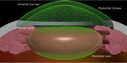
Every eye has a unique size and shape. CATALYS’ advanced 3D imaging technology builds a 3D map of each eye and tailors the treatment to that map. This enables the surgeon to create a customized treatment plan that matches the uniqueness of each eye.

Next, the surgeon uses CATALYS to create a circular opening for accessing and removing the cataract.  Usually, the surgeon does this manually with a sharp instrument and small forceps.  However, the CATALYS machine can create this opening, or capsulorhexis,  for the surgeon using laser.  Clinical studies have shown that this opening is approximately 10 times more accurate when performed with CATALYS than what is achievable by hand.1,2  This opening is an extremely important part of cataract surgery, for it allows the surgeon access to the cataract.  If the opening is not perfectly circular, complications can occur with the placement of the artificial lens.  That’s not to say that an experienced surgeon has to use the CATALYS machine to make the capsulorhexis.  I’ve watched my husband perform cataract surgery numerous times and he has no trouble creating circular opening, however this laser machine does eliminate the chance that opening may be too large or too small, or not perfectly round.  After all, it’s a machine, so it makes the same 5 mm opening every time.  After creating the opening, CATALYS then softens and breaks up the hard cataract into tiny pieces, allowing for gentler, easier cataract removal.3  

Using the manual technique without the CATALYS machine, my husband would have to use the instrument pictured below along with the ultrasound probe to break the cataract into many pieces.

Again, in experienced hands, this technique often results in perfect 20/20 vision, but it does require additional ultrasound power.  With the CATALYS machine, since the laser divides up the cataract, the ultrasound power that is used to remove the cataract is less, which is better for the overall health of the eye.

Depending on the patient’s pre-operative vision and desired visual result, Dr. Wong, or whoever your cataract surgeon is, may recommend a tailored treatment plan that could include creating ultra-precise laser incisions in the cornea and a specific clear lens type, such as a multi-focal lens. This tailored treatment may reduce a patient’s need for glasses or contacts after surgery.

What are the benefits of Laser Cataract surgery?

  • A highly customized procedure using advanced 3D imaging
  • A treatment with little or no discomfort
  • A more precise treatment
  • A gentler and easier cataract removal – this means less trauma to the cornea (the front section of the eye)
  • Generally, a more rapid visual recovery due to reduced inflammation
  • The opportunity to receive tailored treatment with advanced technology multi-focal lenses, which may reduce the need for glasses or contacts after surgery

What is a patient’s experience like with CATALYS ?

Most parts of the surgery are the same with or without the laser.  You still have to check in to the surgical center an hour before the surgery and you can’t eat anything after midnight.  The main difference is the extra step of the laser.  Before going into the operating room where the actual cataract is removed, patients first go into a small room where the laser is located.  The laser machine is pictured below:

Patients like on the bed and the surgeon “docks” the machine on to your operative eye.  Then, he or she will enter the parameters into the machine and guides the machine in making the laser cuts for the capsule opening and sectioning the cataract into pie pieces.  Then, the patient is taken to the operating room, where the surgeon will use the phacoemulsification probe to completely remove the cataract and insert the artificial lens.

A Patient’s view of surgery with the CATALYS

One of the first patients, a nurse at Queen’s, who my husband performed laser cataract surgery on in the summer, was kind enough to record her experience.

Last year, I had the conventional cataract surgery done on my right eye, by Dr Jeff at Queen’s same day surgery. My pre and post op recovery was uneventful, painless, and successful.  This year, for my left eye, I chose to the computerized Optic procedure and the newest Laser Cataract Surgery with CATALYS. Nervous?  No, not at all. I had already researched on line, decided that the Laser Cataract Surgery with CATALYS, would be the most precise and present the fastest recovery. Preoperatively, Dr Jeff thoroughly explained the procedure, and I was ready! The morning of the surgery I arrived at the Surgical Eye Center, was warmly greeted and the preop procedure were started. The Surgical Eye Center has a very relaxed atmosphere. The staff was friendly, informative, very attentive and efficient.  I barely felt the IV start, and appreciated the fact that I did not have to change out of my clothes into a surgical gown. What I was unprepared for, was how painlessly simple the whole procedure would be! I was taken to the room with the Catalys machine, and lay comfortably down on the table.  Dr Jeff explained each step by step of the procedure. I was instructed to focus on the “red light”…and for me this became a light show and a kaleidoscope of red scales and textures. I was mesmerized by the moving lights! After a few seconds (or minutes, I am not sure) a beautiful green light branched out like tiny capillaries, covering the red scales.  Slowly all of the lights and colors faded, and I was assisted to sit up and walked back to the gurney. I was really amazed! I do not remember very much of the second part of the surgery which was the lens implant procedure.  It maybe the effects of the fentanyl or versed, but I think I was still thinking about my “personal light show”. The whole cataract and lens implant took less than 10-15 minutes.I went home, took a tylenol and watched netflix movies! By that evening I was already on the computer checking emails! The next morning I was able to drive to the Honolulu Eye Clinic.Today, I still find myself staring at the leaves of trees and at plants and flowers, as colors are brighter and more vibrant. Post operatively, and I continue to notice that my vision is sharper and I am able to see things more clearly.

The world is really beautiful! This was amazing! Thank you Dr Jeff!

If you are interested in learning more about laser cataract surgery with my husband, Dr. Jeff Wong, in Honolulu, please contact us at Honolulu Eye Clinic at 526-0030 or via email: ronnie@honolulueyeclinic.com and speak with our Surgical Coordinator, Ronnie Bradbury.


Some of you may have noticed I haven’t been posting as much as I normally do.  Those of you who have seen me in person lately know that’s because I’m pregnant!  Actually, now, 37 weeks pregnant and the older I get, each pregnancy gets a little harder.  Though I have gained the least amount of weight of all my pregnancies, I’m still tired and that has left little room for blogging, after work, taking care of two kids and managing our practice.  However, it did remind me that most women are unaware of how pregnancy can actually affect their eyes, so I thought the topic did deserve a post.

Pregnancy is obviously characterized by 40 weeks of hormones from your placenta which now affect the rest of your body.  These hormones can cause physiologic and pathologic changes in Mom and baby.  Also, as a pregnant woman, certain medications are contraindicated if you do develop an eye condition and I wanted to review some of the more common ones.

Common Eye Conditions:

1.  Viral conjunctivitis, or pink eye.  This is characterized by tearing, redness and discharge.  It often spreads from one eye to the other.  You should wash your hands frequently and it is highly contagious the first 5 days of infection. Supportive measures such as cold compresses and artificial tears are the best.  Antibiotic drops are useless against viruses, so no need to take them.

2.  Allergic conjunctivitis – this one drives me crazy, especially in Hawaii during mango season.  You’ll get the same red, watery eyes, but also accompanied by itching and sneezing.  Unfortunately, it’s not recommended to use allergy drops such as Patanol, Pataday or Lastacaft during pregnancy.  So, supportive measures such as cold compresses and artificial tears are the best option during pregnancy.

3.  Dry eyes -Many women experience dry eyes pregnancy due to the hormonal changes.  Symptoms include light sensitivity, feeling like there’s sand in the eyes and even tearing.

Tips

  • Take frequent breaks when reading
  • Use preservative free artificial tears (sensing the theme here? artificial tears are GOOD during pregnancy!).  And, remember – Visine is NOT an artificial tear!

4.  Change in glasses and contact lenses prescription.

You may notice that you don’t seem to be seeing as well with your current glasses or contact lens prescription when pregnant.  Everything becomes fluid filled when you’re pregnant, even your eye structures.  This fluid changes your glasses and contact lens prescription.  Women complain “I can’t see at night”.  Pregnancy can also cause more nearsightedness (myopia) and astigmatism which is temporary.  I always advise pregnant patients not to change their prescriptions when pregnant, because it may not be accurate.  Wait at least few weeks post-partum before getting a new prescription.  If you are nursing, I would wait around 6 months (until you are no longer exclusively breast feeding) to get your new prescription for the highest accuracy.

5.  NO LASIK!!

This goes hand in hand with the recommendation that laser vision correction is NOT recommended during pregnancy or immediately post-partum.  If your prescription is changing, then your doctor cannot be sure of the calculations necessary to give you a good surgical outcome.  Also, the mild sedatives which are given before LASIK and the steroid drops given post-operatively are not allowed during pregnancy.


There are also abnormal eye problems that can occur during pregnancy that you should ‘watch’ out for (i know that was a weak one!).

  • High blood pressure
  • Retinal problems
  • Blood vessel clots
  • Worsening of diabetes
  • Worsening of tumors

1.  Eclampsia – This is very severe high blood pressure after the 20th week of gestation.

It usually occurs with your first child and causes swelling in your legs and problems with kidneys.  It can cause seizures, headaches, stomach pain andvominting as well as vision abnormalities.  This is why your OB will always ask you about headaches or blurry vision.  You might see lightning flashes, blacking out of part of your vision, double vision or it just might be blurry.  Let your OB know right away if you experience any of the above symptoms.  Typically the eyes improve when the medical condition is treated and usually the baby must be delivered to stop eclampsia.

2.  Glaucoma

Some patients have glaucoma and then later become pregnant.  Most of the glaucoma drops are contraindicated in pregnancy because they can induce preterm labor and decrease the baby’s heart rate.  If you have to use drops, then practice punctal occlusion.  That’s when you place the drops in the eye and then hold pressure at the inner corner to minimize the systemic absorption of the drop. 


3.  Gestational diabetes

This is the one I’m blessed with.  I didn’t have it for my first two boys, but now that I’m over 35 years old, my risk increased and I was diagnosed with diabetes.  It has nothing to do with weight gain during pregnancy (I’ve only gained 14 pounds during the entire 37 weeks of pregnancy) or fitness level.  Most people are really surprised to learn that I have gestational diabetes.  Gestational diabetes is when your blood sugar levels become elevated during pregnancy in women who have never had diabetes before. Gestational diabetes starts when the mother’s body is not able to make and use all the insulin it needs during pregnancy. If you have gestational diabetes, you should see your ophthalmologist every trimester to assess for bleeding in the retina.  However, your risk for developing retinopathy is much lower if you were diagnosed with gestational diabetes than if you had a diagnosis of diabetes prior to getting pregnant.  If you had diabetes (Type I or Type 2) before becoming pregnant, than your risk of bleeding in your eyes is higher during pregnancy.   However, the good news is that most long term studies have shown that progression of retinopathy reverses after delivery and that there are no long term changes.

4.  Blood clots in your eyes

As you advance in pregnancy, the blood becomes more viscous and thick.  Clots can form in your legs, lungs and even eyes.  Sudden, profound, painless vision loss must be reported immediately to your OB and ophthalmologist because this could be a “mini-stroke” in your eye or artery occlusion.

5. Central serous chorioretinopathy

This is relatively rare, but I just saw a patient who was in her first trimester who had central serous chorioretinopathy.  Basically, this is a blister of fluid which builds up in the center most part of your retina, called the macula.  Usually these occur in Type A personality (you know, OCD, stressed out, perfectionistic type ) males in their 30’s and 40’s.  It’s induced by stress, hormones, and steroids.  However, those same hormones in pregnancy can cause CSCR in pregnancy.  Patients experience blurry vision, sudden onset of dim vision, waviness in your vision, decreased color vision, and the feeling that things look smaller in one eye than another.  Definitely see your opthhalmologist for any changes in your vision during pregnancy.


Please forgive any typos – I am posting this from the hospital bed where I am in labor!  Finishing this post is helping distract me from the pain of contractions!!


I was listening to the radio the other day about the new concussion policy rules that a lot of youth football leagues now have in place.  It started me thinking about the many eye injuries I see as a consequence of sports and trying to increase awareness about these potentially devastating, completely preventable injuries.  I’m a little late – September was Sports Eye Injury Prevention Awareness Month (that doesn’t exactly roll off the tongue, does it?)  But the message is important, even if the name of the month is a bit long.

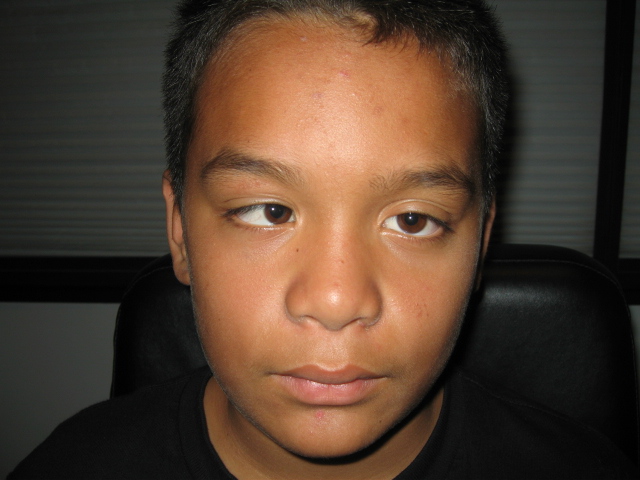
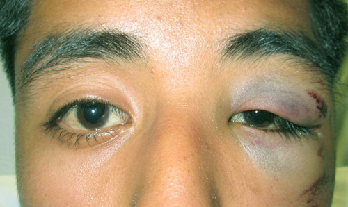
Below is one of my favorite patients and what happened to him should serve as a cautionary tale for all parents whose children are involved in sports.  This kid was 10 years old, playing baseball and accidentally walked behind the batter.  He was struck in the face with the bat (accidentally) and suffered from numerous fractures of his face, the bones around his eyes and lost vision in the right eye from the trauma.

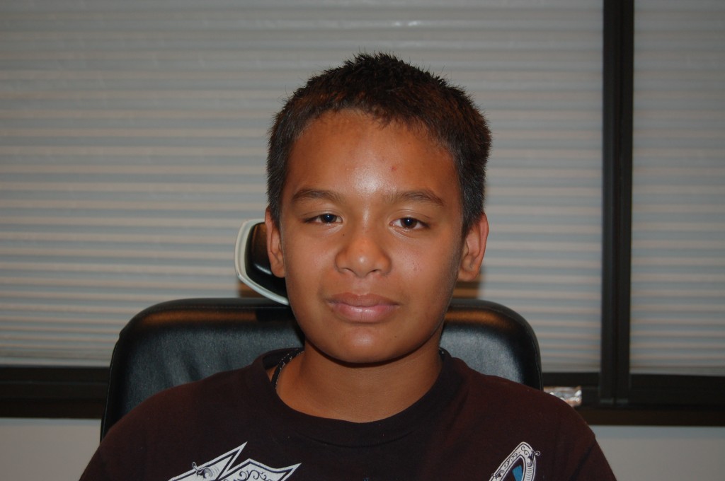
Dr. Timothy McDevitt was able to repair the fractures in Kaliu’s face and I performed surgery to straighten his eyes.  Here he is post-op.  Click here for his mother’s testimonial about her son’s surgical experience at Honolulu Eye Clinic.


He looks great post-operatively and we were able to make the eye look much more normal. Unfortunately, there was no way to restore his vision in the right eye.  As a mother of 2 young boys, this kind of preventable injury hits close to home.  I always think that an injury like this could have been prevented if all of the children on the baseball field had been required to wear sports goggles (also called recreational spectacles).

One problem I’ve noticed is that often kids (and even their parents) will wear their regular glasses when playing sports.  But, you shouldn’t!   More than 40,000 sports-related eye injuries occur every year.  Blunt trauma, such as a ball hitting the eye cause the majority of these injuries.  Some parents also mistakenly think that the helmets with face shields are enough to project the eyes (like football helmets).  But, this is not true.  Your child’s eyes are still exposed to penetrating sports equipment or fingers! Kids and teens have high rates of sports-related eye injuries because of their wide involvement in athletics in school and their tendency to play aggressively. The types of eye injuries can vary including:

1. Corneal abrasions

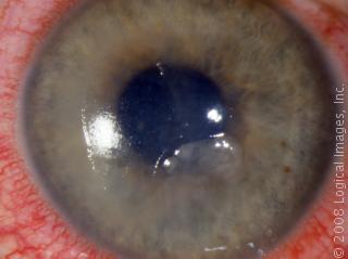
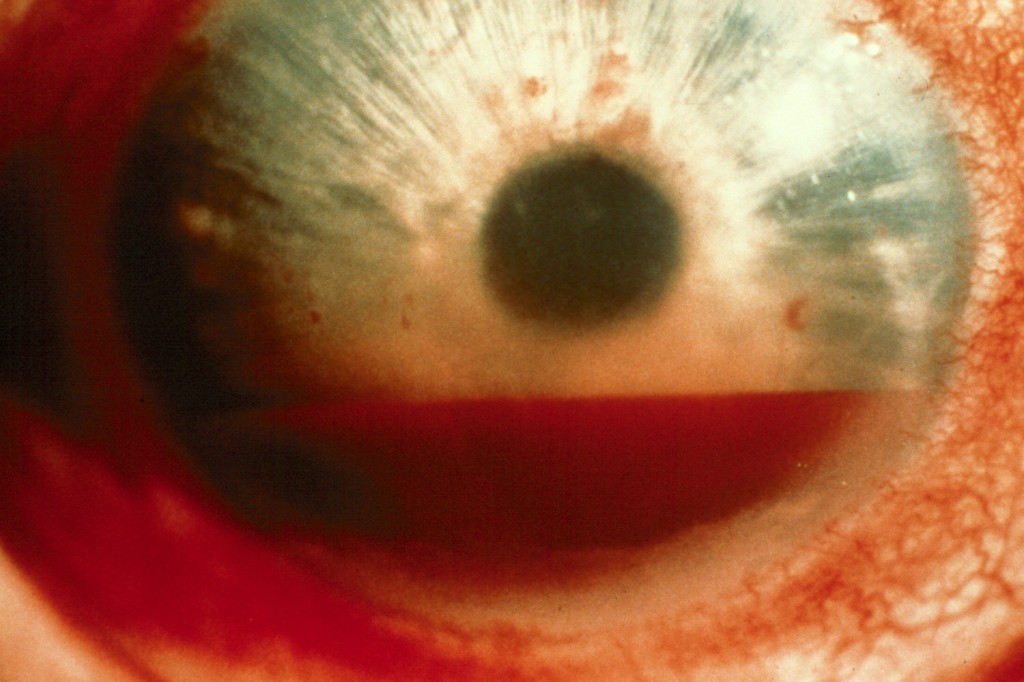
2. Bleeding in the eye (hyphema)

3. Fracture of the bones surrounding the eye (orbital fracture)

4.  Retinal detachment, or even rupture of the eye.


Depending upon which part of the eye is damaged, permanent vision loss can result and surgery may be required to repair the eye. Wearing eye protection significantly reduces the risk of these injuries by ninety percent!

Recommended Protective Eyewear
Many people mistakenly believe that regular glasses or contacts can protect their eyes. Yet, glasses can break upon impact causing a penetrating injury to the eye and contacts do not protect the eye at all.
The American Society for Testing and Materials (ASTM) has standards for the types of sports goggles to be worn in various sports. All sports goggles should be made from polycarbonate.   Polycarbonate is a high-impact resistant plastic that offers UV protection and can be made in prescription or non-prescription lenses. Regular eyeglasses only have 5 percent of the impact resistance of polycarbonate lenses.  Even though all children’s eye glasses are made from polycarbonate, the frames can still bend and break in the eye, making them unsuitable to wear during sports.

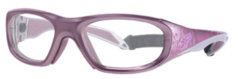
At HEC, we carry Liberty Sport goggles, so I’m most familiar with them, pictured below.

They are a type of protective goggles especially designed to be worn during sports. Fitting most prescriptions, Liberty Sport brand goggles are tested and approved to withstand high impacts. They have very soft padding on the bridge and sides of the frame aimed to protect the bridge of the nose as well as the temples. Their wrap design helps prevent dirt, dust, and even UV rays from damaging the eyeball and the delicate areas surrounding the eye.  Liberty Sport goggles automatically come in the polycarbonate lens material mentioned above and can be ordered clear, or with polarized sunglass lenses, or also with Transitions Lenses (also known as photochromic lenses)–which turn from clear indoors to dark, sunglass-type lens when exposed to UV light outdoors.  They are also designed to fit under sports helmets (baseball, football, etc). And the best part, as mentioned above, is that we can customized them with yours or your child’s glasses prescription in them.  I have some patients that like to wear these as their regular glasses! Of course, they also come in a wide variety of colors and styles.

Whichever type of sports goggles you choose, just make sure that it has polycarbonate lenses and is certified by American Society for Testing and Materials as safety goggles.


Today, we have a guest post by Dr. Jenifer Bossert, staff optometrist, and  head of the Contact Lens Department at Honolulu Eye Clinic.


Sometimes what is old becomes new again!

It happens in fashion, music, and now, even in eyecare.

After 125 years, Scleral Contact Lenses have reemerged as a treatment for certain eye problems.

In 1888 (!), the first scleral contact lens (SCL) was made out of blown glass!  It was very large, 18-20mm, and didn’t allow the eye to breathe so it wasn’t a very healthy. Over time, contact lenses developed from glass, to hard gas permeable, to the soft contact lenses that 89% of our CL wearers enjoy today.  Due to the availability of super-oxygen permeable materials, SCLs are now used to treat a variety of corneal issues.

SCLs are large.  The average gas permeable (hard) lens is 9.5mm.  The average soft contact lens is 14.0-14.5 mm.  SCLs range from 14-22mm.

Below is a picture of a scleral lens on the left and a regular rigid gas permeable lens on the right. Look at that size difference!

SCLs are custom made lenses designed individually for each eye.  Instead of resting on the super-sensitive cornea, they sit on the white part of the eye called the sclera.  Unlike traditional gas permeable lenses, SCLs don’t move when you blink…so, they are very comfortable.

A unique characteristic of SCLs is that the portion of the lens that vaults over the entire cornea is filled with fluid that acts like a liquid bandage to help extremely dry or diseased corneas.  This fluid reservoir also masks corneal irregularities and/or distortions; often, enabling a significant improvement in vision.

Patients who have been told they can’t wear contact lenses may be able to wear a scleral lens. Who is a candidate for the SCLs?  These lenses may be helpful for eyes than are unable to achieve good correction with glasses due to corneal irregularity, those who are contact lens intolerant, or those with significant dryness issues…such as:

  • Kerataconus patients
  • Patients with Sjogren’s Syndrome
  • Corneal transplant patients
  • Severe dry eye patients


So you think you might be a candidate, what next?  In order to begin, we need a current comprehensive eye examination, computerized corneal map, and a scleral contact lens trial fitting.  This is a multi-visit, multi-week process.  There are no SCL manufacturer’s inHawaiiso it’s not unusual for this process to take 8-12 weeks.  And it may take more time to help you master how to apply and remove scleral contacts due to the larger size of the lenses.  In our office, a scleral contact lens training usually takes about 1-2 hours for the first visit and then multiple visits afterwards.

For these reasons, scleral contact lenses cost significantly more than standard contacts; in fact, it is not uncommon for scleral contacts to cost three or four times more.  However, for patients who were previously unable to wear contact lenses at all, these lenses offer a great chance at improved vision.


Anatomy of the eye


You may have friends that say “I have macula.”  What they mean is they have macular degeneration.  Symptoms include:

  • Shadows, blurriness, or holes in the center of vision.
  • Straight lines appear wavy.
  • Trouble seeing details both up close and at a distance.
  • Difficulty telling colors apart, especially ones close in hue.
  • Vision can be slow to come back after bright light exposure.
    People with severe macular degeneration lose their central vision and see the world as depicted below.

In macular degeneration, there is scarring and bleeding in the macula area of the retina.  There  is a spectrum of macular degeneration.  Some patients who have it do not have any visual symptoms at all.  There are two types – dry and wet.  The dry form of macular degeneration, is less severe, yet there’s also no real treatment for it.  It’s considered the early stage of macular degeneration and the only form of treatment is preventative (no smoking, take vitamins such as Ocuvite, which is available at Costco).  The Age-Related Eye Disease Study (AREDS for short) is one of the few studies which proves a beneficial effect of taking a combination of vitamins (Vitamin C, E, beta carotene, zinc and copper) in preventing the progression of macular degeneration.

The wet form, is the more advanced form of macular degeneration.  It’s called “wet” because the blood vessels leak and cause bleeding underneath the macula.

Though this is the worse form of macular degeneration to have, there is treatment for it.

  1. Laser treatment  used to seal off the leaky blood vessels.
  2. Anti-angiogenesis agents – The newest form of treatment are injections (anti-VEGF) which work by slowing the growth of the leaky blood vessels.  Examples of these injections include – Avastin and Lucentis.  Typically, these injections must be performed every 1-2 months or the blood vessels will regrow.

Now to the food part. Some research suggests that a diet rich in those antioxidants may be linked to a lower risk for eye diseases. One recent large study found that people whose diets were high in lutein and zeaxanthin were at lower risk of developing age-related macular degeneration (AMD) and of progression to advanced AMD, the vision-threatening form.  Another large study found that people with high lutein and zeaxanthin dietary intake were at lower risk for cataracts.

Patients always ask if dietary changes will help improve cataracts and macular degeneration.  And, while a healthy diet cannot reverse these changes, it may be able to help prevent macular degeneration and cataracts from becoming significant.  Lutein and zeaxanthin are found in dark, green leafy vegetables like spinach, kale and collard greens. The yolks of eggs also contain lutein. The antioxidants are also present in yellow and orange vegetables and fruits, like sweet potatoes, carrots and peaches. National health organizations recommend from five to 13 servings of vegetables and fruits per day, depending on age and gender. One serving equals one cup of salad greens, or one half a cup cooked vegetables or cut fruit, or one medium-size piece of fruit, or six ounces of juice.

So far, the jury is still out as to whether high dietary levels or supplements of these substances prevent or just slow the progression of age-related eye diseases.  More than one study has shown that vitamins, minerals and phytonutrients found in food offer advantages that are not available in pill form.  Still, eating fresh, whole foods supports a person’s general health, and taking a balanced multivitamin supplement is usually considered a good general health “insurance” practice.

Below are a couple recipes from our family to yours, to maximize your intake of spinach, kale and collard greens.

Now, I am not a huge kale fan, or at least I wasn’t.  But, my mother-in-law made this salad and it fast became one of my favorites.  The recipe is originally from her friend Joan Namkoong.

print recipeKale Salad

Ingredients:

  • Fresh kale
  • Sliced red onion
  • Kalamata or picholine olives, pitted and halved
  • Dried cranberries
  • Walnuts, broken into pieces
  • Balsamic dressing

Directions:

Use a mixture of regular kale, Red Russian kale, lacinato or other varieties and try to get young (small leaves) kale which is more tender.  Remove the leaves from the stems as you wash it; discard stems.  Stack the leaves and cut into fine shreds.  Use about 1 cup of shredded leaves per person.  Place in a salad bowl.

Add the onion, olives, cranberries and walnuts; toss with dressing.  You can toss this ahead of time and let it sit – the kale won’t wilt like other salad greens, making this a great salad for a buffet

Balsamic dressing

Ingredients:

  • ½ cup balsamic vinegar
  • 1 cup olive oil
  • 1 tablespoon honey or sugar
  • 2 teaspoons Dijon mustard
  • 1 teaspoon salt
  • ½ teaspoon black pepper

Directions:

Whisk all the ingredients together

Note:  3:1    oil : acid

Below is a recipe for a popular Indian dish you may have tried in an Indian restaurants.  This recipe is from my mother.

print recipeSaag Paneer (Indian spinach-cheese curry)


Ingredients:

  • 1 bag fresh spinach (frozen spinach can be substituted)
  • 1/2 large onion, chopped
  • 1/2 medium tomato, chopped
  • 2 cloves garlic, chopped
  • 1 inch ginger, finely chopped
  • 1/4 tsp turmeric
  • 1/4 tsp salt
  • 1/2 tsp ground coriander
  • 1/2 tsp ground cumin
  • 1 tsp garam masala
  • 1 Tbsp butter
  • 1 Tbsp oil
  • 1/3 package of paneer, cut into cubes.  (Paneer is simply home made cheese and can be bought frozen in any Indian grocery store, if you don’t want to go through the trouble of making it yourself.  There’s even one here in Hawaii.  Click here for the address.

Directions:

Heat the oil in a large saucepan.  Once warm, add the ginger and garlic and cook for one minute.  Add the onion and reduce the heat to low, cooking for 5 minutes.  Then, add the salt, cumin, coriander, and turmeric to the onion-garlic mixture, cooking for about 2-3 minutes.  Next, add the tomato, cooking it for an additional 2-3 minutes until the tomato begins to soften.  Add garam masala along with 1/4 cup water and cook for 5 minutes.

Add the spinach with 1 cup of water and saute until the spinach begins to wilt, turning off the heat.  Puree the spinach mixture with a hand held blender or remove from heat and blend in a food processor.

In a separate saucepan, heat the butter and paneer, turning constantly so it does not stick to the pan.  Add a pinch of turmeric, garam masala and salt to the paneer while frying.  When golden brown, turn off the heat and combine the paneer with the spinach mixture.


print recipeKale Smoothie (Our 2 year old drinks this!)

Ingredients:

  • 2 stems of kale
  • 1 cup of orange juice
  • 1 cup of frozen peaches
  • 1 cup of frozen pineapples
  • 1 6 ounce cup of yogurt (we use blueberry because it was in the fridge)
  • 5 ice cubes

Directions:

Blend together and serve

And, see, even our 2 year old likes it!


© 2011 Honolulu Eye Doctor & Mom Suffusion theme by Sayontan Sinha